
How it works
NDX has developed a versatile portfolio of cell and tissue acquisition instruments, including UnipicK™, UnipicK+™, and A-picK™, designed for compatibility with a wide range of inverted microscopes.
Our technology is based on an advanced aspiration-based approach that uses precisely controlled vacuum pulses to selectively collect targeted tissue regions or detach and capture adherent cells. Our instrument portfolio ranges from the manually operated UnipicK™ to the fully automated A-picK™, powered by our proprietary PIKCELLS™ software.
Advantages
Our instruments offer exceptional versatility, supporting applications ranging from single-cell collection to precise tissue microdissection. We have also developed specialized protocols for measuring single-cell adhesion forces and selectively acquiring regions of interest from fixed tissue specimens, including formalin-fixed, paraffin-embedded (FFPE) tissues.
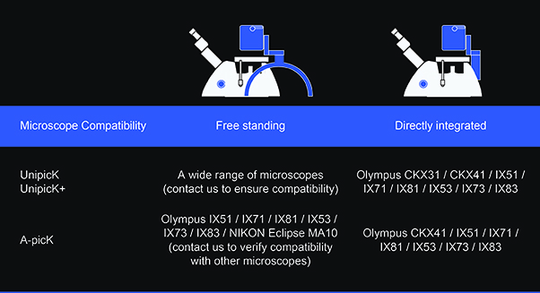
Our instruments enable reliable aspiration and dispensing of volumes ranging from nanoliters to microliters and are compatible with a wide range of inverted microscopes through our proprietary universal microscope straddle.
Both UnipicK+™ and A-picK™ feature intuitive control software with user-friendly menus designed to minimize the learning curve and streamline sample acquisition workflows.
The systems support 96-well plate workflows and are compatible with a broad range of single-cell analysis protocols. Their highly adaptable design allows users to customize acquisition, dispensing, cell detachment, and washing parameters to meet the requirements of specific experimental applications.
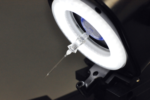
All our instruments support sample collection from standard cell culture dishes and microscope glass slides while requiring minimal consumables. Our proprietary disposable capillary units (DCUs) can also be reused when appropriate, helping reduce consumable expenses and overall experimental costs.
Designed for cost-effective operation, our instruments, including the fully automated A-picK™, offer exceptional value, require minimal routine maintenance, and are backed by a standard one-year comprehensive warranty, with limited warranty coverage available for up to three years.
Summary of advantages
- Cost-efficiency with minimal consumables
- Flexible, fits most inverted microscopes
- High adaptability for customization
- High viability of collected cells
- Versatility in applications
- Functional adhesion test
- Easy to learn and to use
Representative applications
Our instruments support a broad range of applications across cancer and stem cell research, neuroscience, developmental biology, genomics, proteomics, pharmaceutical research, lab-on-a-chip technologies, and fundamental biological research.
Researchers working in fields that require precise isolation of individual cells or selective acquisition of defined tissue regions can benefit from the versatility and precision of our technologies.
Below, we highlight some of the most common and representative applications. For additional examples, please explore our demonstration videos and selected scientific publications.
Single Cell Biology:
- Isolation of single adherent cells from cultures;
- Isolation of rare cells from cultures;
- Isolation of cell clusters or colonies from 3D cultures;
- Clonal expansion of single cells or enrichment;
- Isolation of CTCs from suspensions or placed on a support;
- Isolation of single cells from various biochips;
- Isolation of individual cells from brain sections;
- Measurement of single cell adhesion strength.

Some of the representative publications: Ref1 Ref2 Ref3 Ref4 Ref5 Ref6
Tissue microdissection:
- Microdissection of brain tissues for subanatomical regions and single cell bodies;
- Microdissection of lung, liver, retina, spleen, kidney and other tissues;
- Microdissection of tissue organoids;
- Isolation of regions of interest (ROIs) from fixed tissues including formalin fixed paraffin embedded (FFPE) tissues.
Some of the representative publications: Ref7 Ref8
For information about ordering, delivery, installation, and training, please visit our Frequently Asked Questions (FAQ) page